
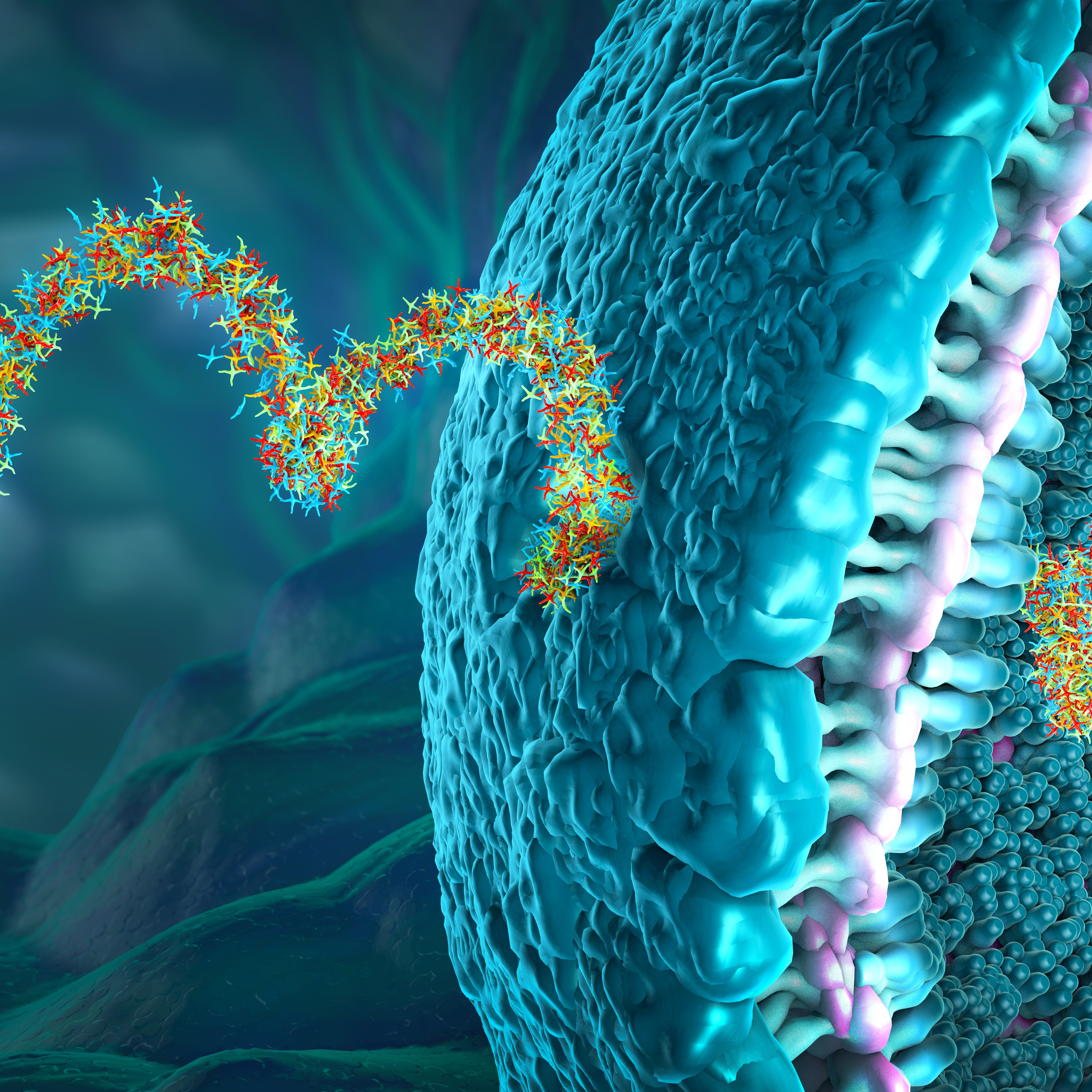
Translatomics for m6A, m3C and ncRNAs
Recent Publications Harnessing the Power of Translatomics
Every week we provide a digest of a small number of recent interesting papers in the field of translatomics.
In this week’s Sunday papers,
- Linder et al. find that mcm5s2U tRNA modification counteracts m6A-dependent mRNA decay.
- Gao et al. develop a novel single nucleotide resolution method m3C-IP-seq for profiling RNA methylomes.
- Liu et al. develop ncPlantDB – a multispecies plant ncRNA database with information about ncRNA-encoded peptides.
tRNA modifications tune m6A-dependent mRNA decay
Cell, 2025
Linder, B., Sharma, P., Wu, J., Birbaumer, T., Eggers, C., Murakami, S., Ott, R.E., Fenzl, K., Vorgerd, H., Erhard, F., Jaffrey, S.R., Leidel S.A. and Steinmetz L.M.
mRNA nucleotides are known to be modified chemically which influences mRNA stability. One of the most frequent mRNA modifications is N6-methyladenosine (m6A). The effects of m6A are thought to be mediated by “reader” proteins that bind m6A sites, e.g. YTH domain family proteins were reported to be involved in the m6A-mediated decay of mRNAs.
With ribosome profiling (Ribo-seq) of monosomes and disomes representing collided ribosomes, researchers discovered that a subset of codons within CDSs are decoded inefficiently when they are m6A-modified, especially at their 3rd sub-codon position. This indicates that m6A reduces codon optimality and thereby marks mRNAs for increased decay. Also, modification of the anticodon loop in tRNA, 5-Methoxycarbonylmethyl-2-thiouridine (mcm5s2U), alleviates this m6A-related effect. This was found by depletion of mcm5s2U which led to stronger translational pausing at m6A-modified codons and more rapid decay of m6A-modified mRNAs. Moreover, by analysing the A-site codon occupancy from Ribo-seq experiments, the authors report that m6A-modified anticodons that are decoded inefficiently lead to ribosome collisions which provides a model that explains m6A-mediated decay that is independent of specific reader proteins.
Besides, mRNAs which demonstrate the coupled m6A and mcm5s2U-dependent decay are enriched in oncogenic signalling pathways and a change this coupling balance may be a cancer biomarker. It has been shown that dysregulation of the m6A and mcm5s2U biogenesis pathways is linked with more aggressive tumors and poor prognosis, and now we have a model that may explain it.
Learn more about EIRNABio’s ribosome profiling services here.
Transcriptome-wide mapping of N3-methylcytidine modification at single-base resolution
Nucleic Acids Research, 2025
Gao, Y., Hou, J., Wei, S., Wu, C., Yan, S., Sheng, J., Zhang, J., Chen, Z. and Gao, X.
One of the prevalent tRNA modifications, 3-Methylcytidine (m3C), has been recently detected in mRNAs, and its precise location and mechanisms of modification in mRNAs have not yet been established. Here the authors developed a novel method called m3C immunoprecipitation and sequencing (m3C-IP-seq) to profile m3C methylomes with single nucleotide resolution. They found modification sites across tRNAs, mRNAs and lncRNAs.
The m3C-IP-seq method uncovered 49 m3C modification sites corresponding to 12 cytoplasmic tRNA isoacceptors and 2 mitochondrial tRNA isoacceptors in HEK293T cells. It also detected 48 high-confidence putative m3C sites in mRNAs and 9 sites in lncRNAs. Interestingly, the majority of m3C sites in mRNAs were enriched within the 3′ UTR region of mRNAs and exhibited a conserved sequence motif, GGACUAC. Further experiments demonstrated the significant increase in the abundance of m3C-modified transcripts within the 3′ UTR following METTL8 knockout compared to non-m3C transcripts. The authors focused on the decay kinetics of the DYNC1H1 mRNA which was confirmed to contain m3C in its 3′ UTR. It was revealed that the half-life of the DYNC1H1 mRNA was significantly extended in METTL8 knockout cells compared to wild-type HEK293T cells. Collectively, the results indicated that the m3C modification in the 3′ UTR of DYNC1H1 promotes its mRNA decay.
Under prolonged hypoxia conditions, the authors noticed a reduction in METTL8 expression in HEK293T which resulted in a progressive decrease in m3C methylation in mRNAs. In summary, m3C modification in the 3′ UTR contributes to the degradation of m3C-modified mRNAs, linking this epitranscriptomic mark to mRNA turnover.
ncPlantDB: a plant ncRNA database with potential ncPEP information and cell type-specific interaction
Nuclear Acid Research, 2025
Liu, L., Liu, E., Hu, Y., Li, S., Zhang, S., Chao, H., Hu, Y., Zhu, Y., Chen, Y., Xie, L., Shen, Y., Wu L. and Chen M.

In recent years, annotated non-coding protein coding RNAs (ncRNAs) have been shown to play a regulatory role in gene expression during plant development and stress responses. Increasingly, many ncRNAs have also been found to have protein-coding potential resulting in ncRNA-encoded peptides (ncPEPs) which have been shown to play important roles in plant biology and agriculture. To aggregate the community knowledge, the authors developed ncPlantDB, a comprehensive database integrating ncRNA and ncPEP data across 43 plant species which combines 353,140 ncRNAs, 3799 ncPEPs and 4,647,071 interactions.
The authors manually reviewed over 300 publications to identify 91 ncPEPs (51 experimentally validated) across multiple plant species. To further validate ncRNAs’ coding potential, the researchers collected ribosome profiling (Ribo-seq) data from the NCBI SRA database for Arabidopsis thaliana, Oryza sativa, Solanum lycopersicum, Zea mays and Triticum aestivum. Ribo-seq data were uniformly reprocessed and unannotated translated regions (translons) were detected using the Ribocode tool resulting in 743 small ORFs predictions. In addition, single cell RNA-seq datasets were analysed to develop comprehensive cell-specific co-expression networks and identify the regulatory roles of plant ncRNAs. Information on the coding potential of circRNAs was sourced from PlantcircBase which led to a collection of 2 965 putative ncPEPs.
Altogether this resource provides unique characteristics including cell-specific interaction networks derived from single-cell RNA sequencing and translation data derived from Ribo-seq and the literature.
Learn more about EIRNABio’s ribosome profiling services here.